 English
 中文
中文 ဗမာစာ
ဗမာစာ
Retinal veins are blood vessels in the retina (nerve layer) of the eye that drains blood from the eye. If retinal veins are occluded (blocked), blood and fluid leak into the surrounding retina. This can be likened to a dam causing a river to burst its banks, resulting in flooding and damage to the surrounding land.
When leakage occurs into the central fixation point of the eye (macula), swelling of the macula (macular edema) will cause visual impairment. Sometimes, vein occlusions can also cause impediment of blood flow into the eye, and this poor blood supply can lead to other complications like severe visual loss, growth of abnormal blood vessels and high eye pressure (glaucoma).
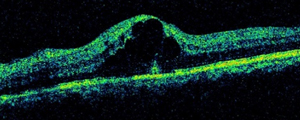
A scan of the macula shows severe swelling due to the vein occlusion. Central vision
can be severely affected and should be treated early to prevent permanent damage.
There are 2 types of vein occlusions.
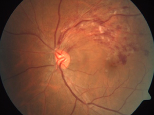
BRVO
A branch retinal vein occlusion (BRVO) is due to blockage of one of 4 large retinal veins, each supplying a quarter of the retina.
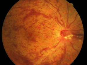
CRVO
A central retinal vein occlusion (CRVO) is due to blockage of the main vein supplying the entire retina. In general, a CRVO is more serious than a BRVO.
Causes
The exact cause of vein occlusions is not known but there are some risk factors. These include increasing age, high blood pressure, diabetes, high cholesterol levels and glaucoma. Smokers and individuals with certain blood disorders are also more likely to get vein occlusions.
Treatment
If the occlusion causes macular edema, intravitreal injection therapy may be used to reduce the swelling and reduce damage to the central fixation point of the eye. This involves the injection of medication (antiVEGF agents or steroids) into the eye using a very tiny needle, and has been shown in clinical studies to deliver good vision results. The injection is done as an outpatient procedure and multiple injections may be necessary.
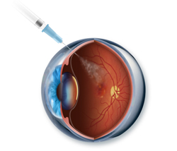
Drugs are delivered directly into the eye for maximum effect.
In some cases, laser therapy is done to treat areas that are severely affected by poor blood supply. The purpose of laser therapy in these cases would be to reduce the risk of complications like more severe bleeding and glaucoma.
Laser therapy is done as an outpatient procedure and may need to be performed over a few sessions.